

An artificial aortic valve must open with the flow of blood, allow left ventricular ejection while maintaining a controlled pressure gradient, and close while minimising regurgitation. It performs this cycle tens of millions of times each year, within an environment where materials, geometry, blood flow and biological tissues interact continuously.
Since the first prosthetic valves were implanted in humans, valve engineering has progressively incorporated new design constraints: material haemocompatibility, haemodynamic performance, durability, minimally invasive implantation, advanced imaging and patient-specific planning.
With Transcatheter aortic valve implantation, TAVI, the prosthesis has become inseparable from its delivery system, the imaging used to plan its deployment and the anatomy in which it must function.
The next phase centres on durability, reintervention and management of the device across the patient’s lifetime.
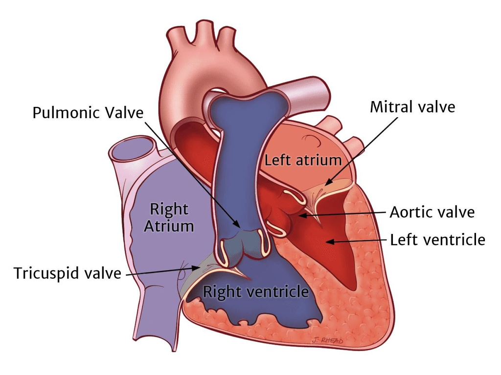
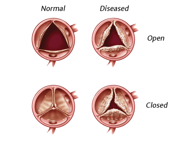
Left: heart chambers and valves — cardiac anatomy and the position of the aortic valve at the left ventricular outflow.
Right: illustration of a normal aortic valve and a calcified, stenotic aortic valve. © Edwards Lifesciences
From Mechanical Prostheses to Living Tissue
The First Mechanical Valves
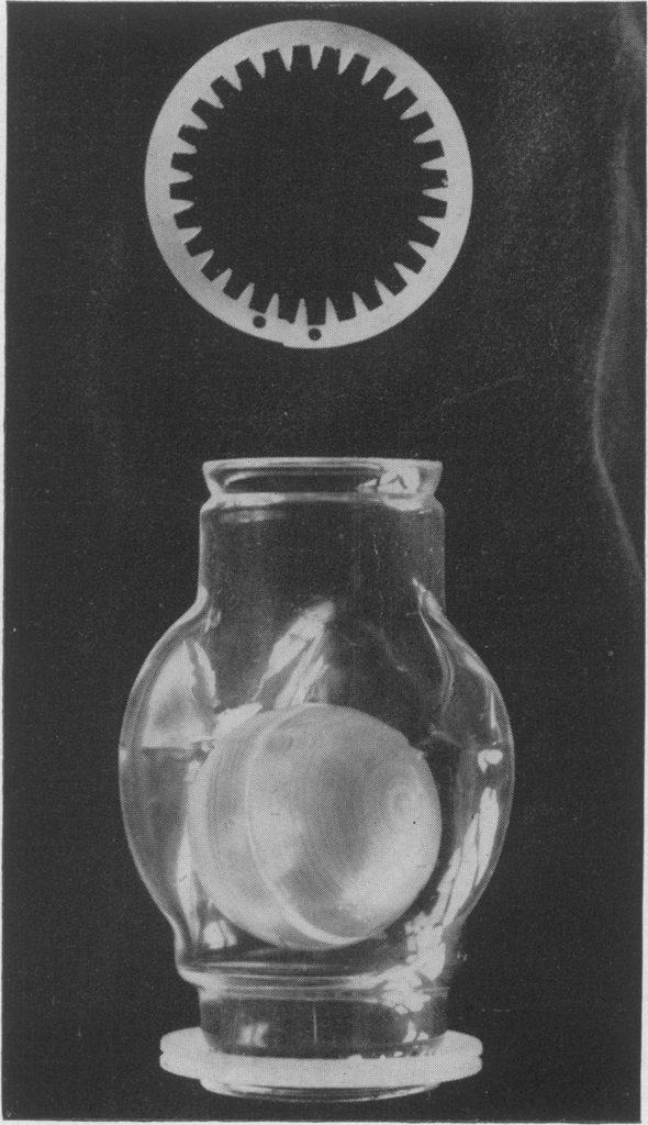
On 11 September 1952, Charles Hufnagel implanted a ball-valve prosthesis in the descending thoracic aorta of a patient with aortic regurgitation. The device did not anatomically replace the native valve, but demonstrated that a mechanical prosthesis could provide effective valvular function in humans. The milestone is documented in the history of the first prosthetic heart valves.
Hufnagel valve (1952), one of the pioneering devices in modern valve engineering. © Postgraduate Medical Journal / BMJ — 1956
The advent of cardiopulmonary bypass subsequently made direct valve replacement possible. In 1960, Dwight Harken performed an orthotopic aortic valve replacement using a ball-and-cage mechanical prosthesis. That same year, Albert Starr and engineer M. Lowell Edwards developed the Starr-Edwards valve, whose evolution is documented in historical accounts of mechanical heart valves.
Before the advent of TAVI, aortic valve replacement was primarily performed through open-heart surgery, involving sternotomy and cardiopulmonary bypass.
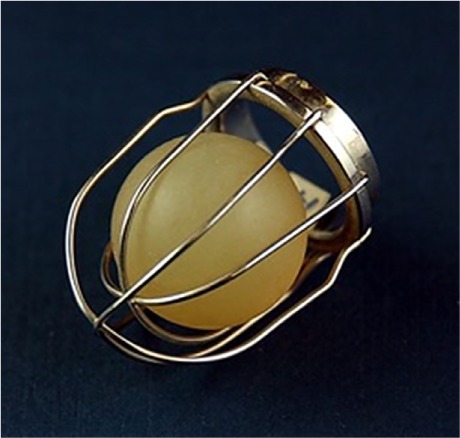
Harken-Soroff valve (1960) — double stainless-steel cage and silicone ball, designed by Dwight Harken for aortic valve replacement. © Museum of Medical History, Sierra Sacramento Valley Medical Society & Bioengineering Department, California State University, Sacramento
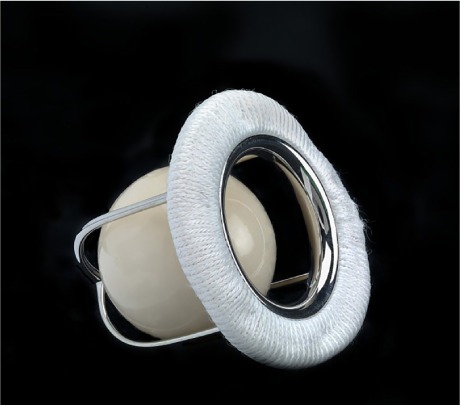
Starr-Edwards valve (1960) — metal cage and Silastic ball, characteristic of the first generation of mechanical prosthetic valves. © National Museum of American History, Smithsonian Institution, Washington, DC
Later generations evolved from single-disc to bileaflet designs. Their architectures evolved alongside their materials: metal alloys, pyrolytic carbon and coatings were selected for their mechanical properties, fatigue resistance and interaction with blood.
Mechanical valves remain highly durable, although lifelong anticoagulation is required for most patients to reduce the risk of thromboembolism.
Tissue Enters Valve Engineering
Bioprosthetic valves use treated and stabilised porcine tissue or bovine pericardium to form the leaflets. At Abbott, the Epic™ range exemplifies this approach, using porcine tissue in a design focused on calcification resistance and durability.
The development of bioprostheses introduced another variable: biological ageing. Calcification, mechanical fatigue, leaflet tears and structural valve deterioration can progressively alter the properties of implanted tissue. The mechanisms underlying the deterioration of biological replacement valves have been studied for decades.
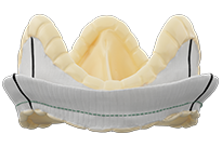
Epic™ valve — surgical bioprosthesis with porcine leaflets developed by Abbott, designed with potential future intervention in mind. © Abbott
From Open-Heart Surgery to the Catheter
The Prosthesis Enters the Arterial Pathway
In the early 1990s, research into transcatheter implantation led to a new configuration: a bioprosthetic valve mounted on a metal frame, crimped onto a delivery system and advanced through the vasculature to the native valve. The transfemoral approach subsequently became the principal access route for TAVI.
On 16 April 2002, in Rouen, Professor Alain Cribier performed the first human implantation of a transcatheter aortic valve in a patient with severe aortic stenosis who was considered inoperable. The procedure is now regarded as the clinical starting point of modern TAVI.
The prosthesis had to meet several requirements simultaneously: a low insertion profile, resistance to compression, controlled deployment, secure anchoring within a frequently calcified native valve, stability and haemodynamic performance.
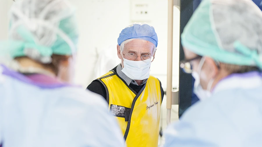
Professor Alain Cribier (1945–2024), pioneer of percutaneous treatment for valvular disease and a founding figure in TAVI. Since then, more than four million patients worldwide have been treated with TAVI. © Institut Alain Cribier
Anatomy Becomes a Design Input
Cardiac CT characterises the aortic annulus, aortic root, calcification, coronary ostial height and access routes. These measurements determine prosthesis sizing and position, as well as the feasibility of implantation. The 2025 ESC/EACTS recommendations on valvular heart disease incorporate cardiac CT into the selection and planning of valvular procedures.
Geometry Meets Fluid Mechanics
Effective orifice area, transvalvular gradients, flow velocities, regurgitation and prosthesis–patient mismatch (PPM) are key parameters in assessing prosthetic valve function.
Geometry also influences flow around the leaflets, implant stability and the conditions for potential future coronary access.
The Valve as a Technology Platform
Edwards: Materials, Valve Design and Durability
Edwards Lifesciences occupies a central position in TAVI.
The Monaco Cardiothoracic Centre (CCM), Siemens Healthineers’ leading reference centre for cardiovascular medicine, uses, among others, Edwards SAPIEN 3 Ultra.
The SAPIEN 3 Ultra RESILIA extends this R&D approach through a technology designed to preserve pericardial tissue and limit calcification. It reflects the evolution of valve design towards biomaterial durability.
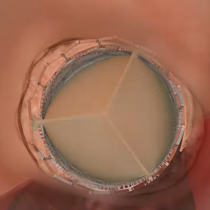
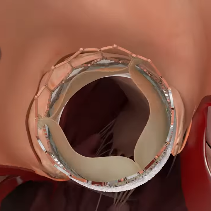
SAPIEN 3 Ultra RESILIA — Transfemoral Procedure © Edwards Lifesciences
In 2026, Edwards is pursuing this trajectory through the PROGRESS trial, which is evaluating SAPIEN valves in patients with moderate calcific aortic stenosis.
Valvular R&D now focuses as much on how tissue behaves over time as on implantation performance.
Infrastructure as an Integral Part of the Device
The Hybrid Theatre
The development of TAVI brings imaging, catheterisation, surgery and delivery systems together within a single environment.
The CCM has two hybrid theatres and a Naeotom Alpha® photon-counting CT scanner from Siemens Healthineers, dedicated exclusively to cardiovascular imaging. The CCM was the first private centre in the world to install a photon-counting CT scanner of this type.
Acquisition, reconstruction and interpretation of anatomical information are integrated into the same interventional pathway.
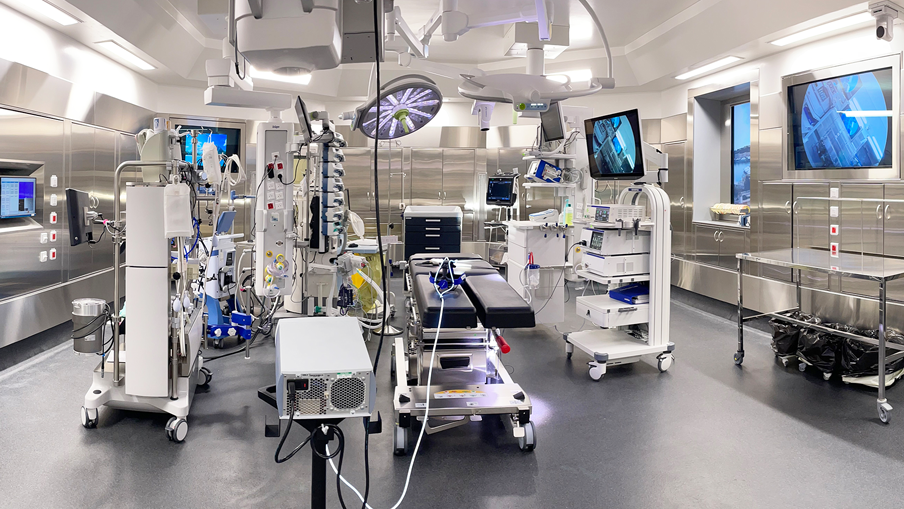
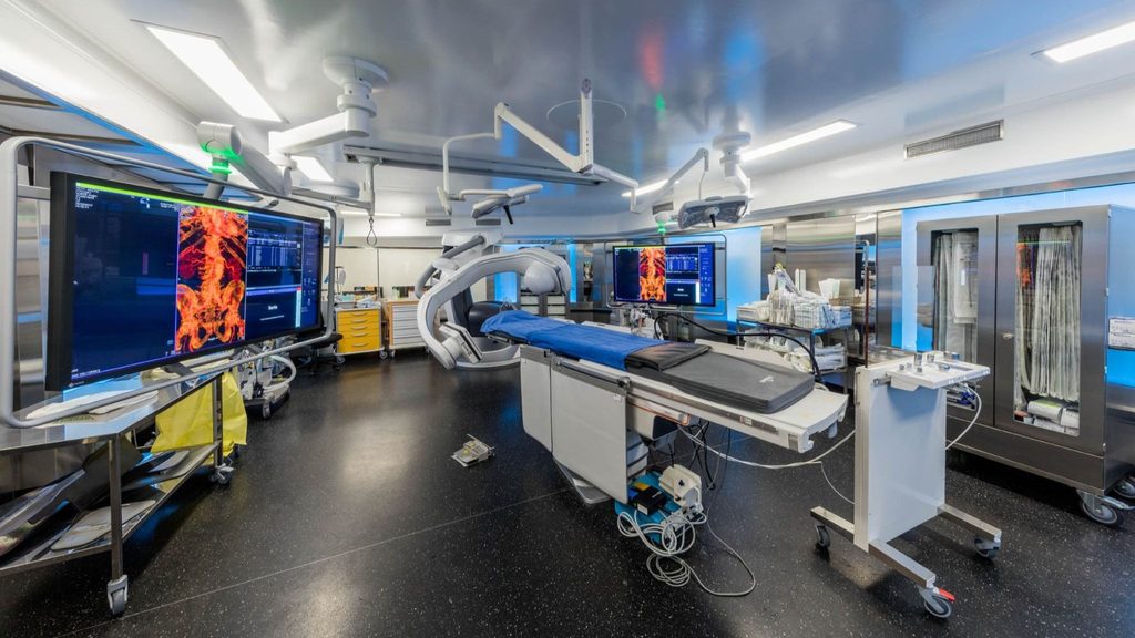
CCM hybrid theatres, equipped notably with the ARTIS pheno et ARTIS icono interventional angiography systems from Siemens Healthineers, integrating advanced imaging into the interventional procedure. © CCM
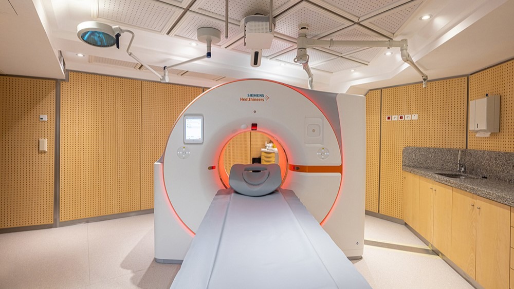
Naeotom Alpha® photon-counting CT scanner from Siemens Healthineers, dedicated to cardiovascular imaging at the CCM. The centre was the first private institution in the world to be equipped with this technology for the specialty. © CCM
The delivery system
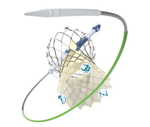
In TAVI, prosthesis efficacy also depends on how precisely it can be delivered, positioned and deployed within the patient’s anatomy.
Medtronic addresses this through the Evolut™ FX system, integrating the bioprosthetic valve with a delivery system designed for controlled navigation, deployment and positioning. The Confida™ Brecker Guidewire, développé spécifiquement pour les procédures TAVI, complète cette chaîne d’accès et de support.
Evolut™ FX — transcatheter aortic valve and delivery system. © Medtronic
From CT to the Digital Model
Computational modelling is increasingly being used to reproduce interactions between the prosthesis, tissues and blood flow before intervention.
A multidisciplinary review published in 2026 in European Heart Journal – Digital Health examines the use of such models to study TAVI durability. Simulations can reproduce prosthesis deployment, mechanical stresses, stent deformation and flow patterns that may contribute to structural deterioration. The nevertheless emphasises that these predictions remain exploratory: validation linking simulated markers to long-term clinical outcomes has yet to be established.
AI Enters Procedural Planning
Artificial intelligence (AI) is increasingly being used to automate the analysis of pre-TAVI CT scans, including segmentation of the annulus and aortic root, measurement of dimensions and coronary ostial distances, and assessment of calcification. These tools can accelerate and standardise prosthesis sizing and procedural planning.
Circle Cardiovascular Imaging, through its cvi42, platform, offers automated measurements as well as virtual device visualisation and femoral access planning.
From imaging to planning: cvi42 converts cardiac CT data into measurements for TAVI planning. © Circle Cardiovascular Imaging
Valve durability
An Implant Shapes the Therapeutic Pathway
The recommandations of the European Society of Cardiology (ESC) and the European Association for Cardio-Thoracic Surgery (EACTS), published in 2025, incorporate age, life expectancy, anatomy, durability and reintervention options into the choice of valvular strategy. In patients aged 70 years or older with tricuspid aortic stenosis, TAVI is recommended when anatomy is suitable; in younger patients, surgery retains an important role when several interventions may be required over the course of a lifetime.
The choice of the first prosthesis therefore shapes what follows: coronary access, risk of prosthesis–patient mismatch, positioning, geometry and the feasibility of future reintervention. Lifetime management means viewing the prosthesis not as a definitive device, but as the first element in a therapeutic pathway.
Very long-term data remain more limited for transcatheter valves, particularly in younger patients, in bicuspid valves and in valve-in-valve procedures. Any subsequent intervention will depend, among other factors, on the geometry of the initial prosthesis, commissural alignment, its relationship with the coronary arteries and the space available for a potential TAVI-in-TAVI procedure.
From Valve to Cardiovascular System
A valve’s performance depends on the interaction between materials, geometry, haemodynamics, anatomy, imaging, delivery systems and clinical data.
Design now extends beyond the prosthesis itself to the technological ecosystem in which it is planned, delivered, monitored and, ultimately, replaced.
The World Heart Vision 2030 identifies innovation and technology as key drivers of future cardiovascular health. The aortic valve offers a tangible example of this in practice.

“Don’t miss a beat” with Cardi V.
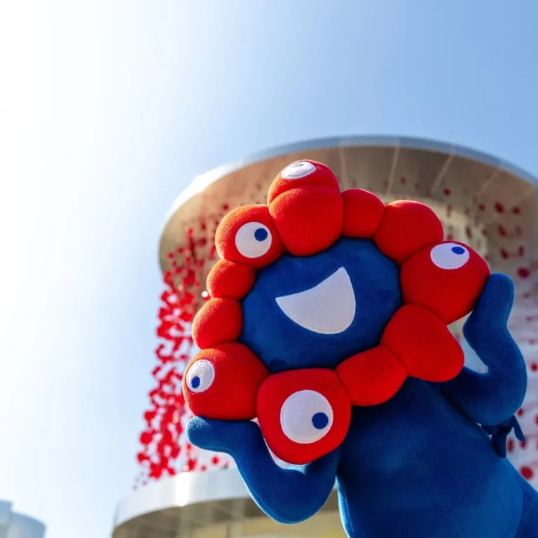
Cardi V, official mascot of the World Heart Day 2026 campaign, 29 September © World Heart Federation